
UC San Diego Professor Mark Ellisman Presents Neuroscience Research Findings to Emperor of Japan
Sapporo, Japan and San Diego, CA, September 6, 2006 -- A pioneer in the development and use of new bioimaging tools for neuroscience is one of only three experts -- and the only non-Japanese national -- invited to outline their research to Japan's Emperor Akihito and Empress Michiko when they visited the 16th International Microscopy Congress in Sapporo, Japan today.
|
“Professor Ellisman has pursued an intensely interdisciplinary approach to answering open questions about the brain’s function and structure,” said UC San Diego Chancellor Marye Anne Fox . “This honor bestowed by the Emperor of Japan is also in recognition of Dr. Ellisman’s and UCSD’s track record of reaching out to foreign partners.”
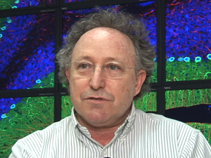
“This honor has particular resonance for me because we have collaborated with scientific groups and companies in Japan for over twenty years,” said Ellisman. “Our joint work with advanced 3D electron microscopy is helping us to understand the normal and abnormal structure and function of the nervous system. There are no modern high-voltage electron microscopes available to us for basic biological and biomedical research in the United States . We are very thankful to the Japanese government for enabling our access to these high-powered instruments for collaborative projects.”
Emperor Akihito -- a biologist and ichthyologist who still publishes research papers -- asked many questions, and thanked Ellisman for his work to foster more international collaboration.In his presentation to the Emperor, Ellisman stressed the value of observing the brain at multiple scales—from the very large (gross anatomical) to the very small (molecular) level. “Imagine looking at the brain as if you were looking at the globe using Google Earth,” he noted. “Zoom out and you get the big picture. But you may also want to zoom in to a particular mountain formation, and zoom in even further to observe a specific volcano. Visualizing the data at multiple scales provides insights that are just not possible if you only look at one image—and that’s true whether you’re looking at the Earth or the brain.”
In Sapporo, Ellisman showcased three ‘multi-scale’ projects that have benefited from advances in high-energy electron microscopy and electron tomography. All three projects are based in organizations that Ellisman directs, including the UCSD-based National Center for Microscopy and Imaging Research (NCMIR), and the Biomedical Informatics Research Network Coordinating Center , funded by the National Institutes of Health (NIH) National Center for Research Resources.
Using electron tomography, for example, Ellisman and his team were able to determine the precise geometry of synapse structures—leading to simulations of physiology that are lending new insights into scientists’ understanding of synaptic transmission.
|
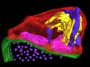
NCMIR also developed tools that allow researchers to switch from viewing whole 3D cells under the light microscope to viewing electron microscopy of photo-oxidized 3D cells. Adding electron tomography capabilities to Japan’s largest high-voltage electron microscope allowed Ellisman’s team to look through very thick sections (5-10um) and obtain very accurate views of dendritic spines—the bottle brush-like protrusions of neuronal dendrites that receive 90 percent of the synaptic connections in the mammalian brain (see image above).
With his collaborators, Ellisman is studying disorders that involve synapses, such as mental retardation resulting from Fragile X syndrome (the primary inherited cause of mental retardation). They observed these spines in normal mice and in strains of mice genetically modified to represent a form of human mental retardation. Preliminary results show that neurons in the cerebral cortex of mice exhibit altered dendritic morphology when the Fragile X mental retardation protein gene FMR-1 is removed.
“This is a possible mechanism contributing to the disordered brain microstructure that may lead to mental abnormalities,” concluded Ellisman. “These observations help us understand what goes wrong with the wiring of the brain that results in learning deficits, and these findings may have broader implications for the underlying mechanisms of learning and memory or perhaps reveal the mechanisms which underlie autism.”
|
Simultaneously, Ellisman is working within the California Institute for Telecommunications and Information Technology’s OptIPuter project to allow scientists in San Diego to “see” what the Osaka microscope sees, in real time and in high-definition video over dedicated optical networks. His work on that project is funded by the National Science Foundation.
Professor Mark Ellisman joined the UCSD faculty in 1977 after receiving his Ph.D. in molecular, cellular and developmental biology from the University of Colorado , Boulder . He established NCMIR in 1988 to achieve greater understanding of the structure and function of the nervous system by developing 3D light and electron microscopy methods. A founding fellow of the American Institute of Medical and Biological Engineering, Ellisman has served as the founding director of the UCSD Center for Research in Biological Systems since 1996.
Related Links
16th International Microscopy Congress
National Center for Microscopy and Imaging Research
Biomedical Informatics Research Network-Coordinating Center
National Institute of Neurological Disorders and Stroke
National Institute for Research Resources
National Institute for Physiological Sciences (Japan)
Center for Research in Biological Systems
Media Contacts
Media Contact: Doug Ramsey, (858) 822-5825, dramsey@ucsd.edu