

NCMIR Wins Prestigious NIH Scientific Visualization Competition
By Stephanie Sides / NCMIR
San Diego, October 17, 2016 — The National Center for Microscopy and Imaging Research (NCMIR) has a long-established reputation based on addressing important biomedical research challenges, the ever-more-demanding requirements of big datasets, and how to explain results in an understandable yet compelling way using scientific visualization. On October 13, all three areas of expertise were recognized when the National Institute of General Medical Sciences (NIGMS) was awarded first place for its NCMIR-submitted visualization in the 2016 Combined Federal Campaign NIH Institute and Center Directors’ Art Challenge for “The Beauty of Science”.
Each year NIH Institute and Center (IC) directors are invited to participate in a “Directors’ Challenge” to increase institute awareness, create excitement about the programs they fund, and inspire donations to the Combined Federal Campaign. This year’s challenge, led by the National Institute on Drug Abuse, was an art competition. Each IC director was invited to submit his or her organization’s best work of art depicting the “Beauty of Science.”
NCMIR submitted multiple entries to NIGMS; two were recognized with first and second place awards. NIGMS, in turn, submitted NCMIR’s first place selection to the general competition, and it was that image that won overall.
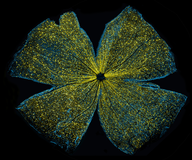
The winning image, titled “Lighting Up the Promise of Gene Therapy for Glaucoma,” shows what looks like the gossamer wings of a butterfly. But this image, sparkling with fluorescent molecules and delicately snipped to lay flat, actually shows the retina of a mouse.
It was selected by combined voting of the NIH IC directors, each of whom cast votes for their three favorite entries. The winning artwork will be displayed in Building 1 at NIH, on the Combined Federal Campaign website, and in future NIH publications.
This image originally accompanied the peer-reviewed paper published in Cell Death & Disease, cited below. In announcing the award in an e-mail to the NCMIR team, Dr. Paul Sammak, Program Director in the Division of Biomedical Technology, Bioinformatics and Computational Biology at NIGMS, referred to the research as “elegant work that describes the encouraging story about the promise of therapy for glaucoma from basic science studies.”
Keunyoung {Christine) Kim, first author on the paper and creator of the image, says, “It’s amazing to see such intricate and artistically organized microscopic structures. I encountered an entirely new world invisible to the naked eye.” She performed light microscopy, serial blockface electron microscopy (known as SBEM), and analysis related to mice retina and the optic nerve head. She also used ImageJ Mosaic plugins, developed at NCMIR, and IMOD, developed at the University of Colorado, Boulder.
About the Science
The term glaucoma represents a group of eye diseases that lead to progressive optic nerve degeneration, resulting in visual field loss and irreversible blindness. It is characterized by the death of neurons in the retina called retinal ganglion cells (RGCs). Several studies over the past decade have suggested that targeting these cells with gene therapy designed to prevent their death might slow the progression of glaucoma. As a result, when Kim works on projects focusing on vision protection in glaucoma, she typically works with RGCs.
Kim explains that, “RGCs are a population of neurons in the central nervous system that affect the transportation of visual information from the eye to the brain and are a key element in the pathophysiology of all forms of glaucoma.” She adds that the number, distribution, and morphology of RGCs vary in central and peripheral areas. So, mapping the entire retinal structure is key to elucidating RGC function. By imaging large regions of whole retinas at high resolution using large-scale confocal light microscopy and mosaic imaging, a combined technique pioneered by NCMIR, she was able to characterize RGC distribution from central to peripheral retinal areas. “It’s similar to Google Earth in that we computationally stitch together many, many small high-resolution images,” said Ellisman, who also directs the Center for Research in Biological Systems, which promotes cross-disciplinary research involving NCMIR, the San Diego Supercomputer Center, the California Institute for Telecommunications and Information Technology (Calit2) and its Qualcomm Institute division at UC San Diego, and UC San Diego Health Sciences.
Kim and colleagues used a particular type of virus system to deliver therapeutic genes into the mouse retina. More specifically, they introduced into the virus a gene for green fluorescent protein (GFP) to visualize how well the virus transduced the cells. Two months after viral delivery of the gene to the eyes of seven-month-old mice, the researchers examined the retinas of the subjects. They observed GFP expression (yellow) in all parts of the retinal ganglion cells, including the soma, axons, and dendritic tree. Significantly, these results suggest that such a viral delivery system could deliver therapeutic genes to retinal ganglion cells to treat glaucoma and related diseases.
Citation: Kim, KY, GA Perkins, MS Shim, E Bushong, N Alcasid, S Ju, MH Ellisman, RN Weinreb, and WK Ju, "DRP1 inhibition rescues retinal ganglion cells and their axons by preserving mitochondrial integrity in a mouse model of glaucoma." Cell Death & Disease 6, No. 8 (2015): e1839.
Related Links