New Technique to Study How Proteins and Ligands Interact
San Diego, December 6, 2016 — A team of researchers has developed a more accurate and less disruptive method to study how proteins and the small molecules that bind to them, known as ligands, interact. The method, called Transient Induced Molecular Electronic Spectroscopy (TIMES), could be used as a tool to better understand protein chemistry and to accelerate drug discovery and development.

“This new tool will enable researchers to investigate a larger variety of chemical and biochemical reactions than was previously possible,” said Yuhwa Lo, electrical engineering professor at the University of California San Diego and senior author of the study. The work, a collaboration between UC San Diego and Lamar University in Texas, was published recently in the journal ACS Central Science. Lo also directs the Qualcomm Institute's Nano3 nanofabrication cleanroom facility on the UC San Diego campus, as well as the newly NSF-funded San Diego Nanotechnology Infrastructure (SDNI).
Protein-ligand interactions are of particular interest to biopharmaceutical researchers because of their applications in drug discovery. During the early stage of drug discovery, researchers screen millions of small molecule drug candidates, such as ligands, in order to find the ones that can most effectively combine with a target protein.
But one of the biggest challenges in early drug discovery is that there is currently no good way to detect protein-ligand interactions without requiring chemical or structural modifications, Lo explained. These modifications can affect protein-ligand binding, potentially skewing the measurement, he said.
Lo and colleagues developed TIMES, a new high-resolution imaging technique that can accurately characterize protein-ligand interactions without perturbing their binding. In the TIMES technique, proteins, ligands and protein-ligand complexes flow through a device containing an electrode surface connected to a low-noise electric amplifier. As molecules approach the electrode surface, electric signals are produced. The signals can be analyzed to determine exactly how proteins and ligands bind.
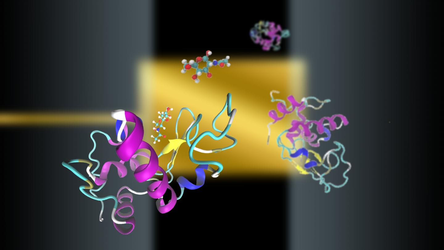
Using TIMES, researchers were able to accurately measure the interactions between a protein called lysozyme with a ligand called N-acetyl-D-glucosamine (NAG) and its trimer, NAG3. While NAG and NAG3 are relatively similar in size and chemical makeup, experiments showed that NAG3 had almost a thousand-fold greater binding affinity to lysozyme than NAG, which agreed well with results published in the literature.
“That’s another advantage of this method. It can actually differentiate the degree of binding — for example, what percentage improvement in binding — between similar molecules. Current methods typically just tell you whether or not binding occurs,” Lo said.
An area of improvement with the TIMES method involves increasing the sensitivity of the technique. “We’re working towards being able to do single molecule detection with this method,” Lo said.
###
Full paper: “Protein-Ligand Interaction Detection with a Novel Method of Transient Induced Molecular Electronic Spectroscopy (TIMES): Experimental and Theoretical Studies.” Authors of the study are: Tiantian Zhang, Yuanyuan Han, Ping-Wei Chen and Yu-Hwa Lo of UC San Diego; and Tao Wei, Heng Ma, Mohammadreza Samieegohar and Ian Lian of Lamar University.
This research is funded in part by the National Science Foundation (grant ECCS-1610516) and Vertex Pharmaceuticals, Inc.
Media Contacts
Liezel Labios, (858) 246-1124, llabios@ucsd.edu
Related Links

